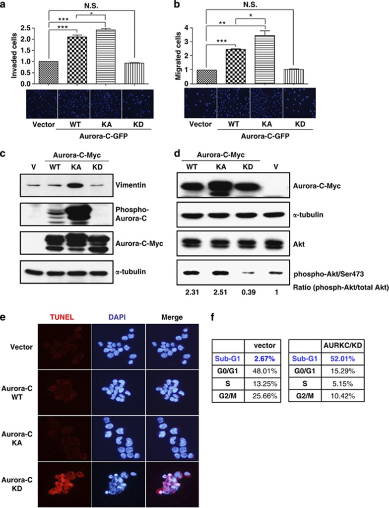
Figure 7.
Aurora-C-GFP enhances cell migration, invasiveness, and survival in a kinase-dependent manner. (a and b) Equal numbers of cells that expressed Aurora-C-GFP/WT, KA, or KD were seeded onto collagen-coated (a) or matrigel-coated (b) transwell inserts. Invasive or migrated cells were counted after 24 h of incubation and subsequent staining with DAPI. The quantitative results of three independent experiments are shown. *P<0.05; **P<0.01; ***P<0.001. NS, not significant. (c) HeLa cells expressing Aurora-C-Myc/WT, KA, or KD were used for IB. The expression levels of vimentin, Aurora-C, and phospho-Aurora-C are shown. Vector (V)-transfected cells were used as a background control. (d) Total lysates of HeLa cells with Aurora-C-Myc/WT, KA or KD expression were prepared to determine the expression levels of phospho-AKT (phosphor-Akt/Ser473), AKT, and Aurora-C-Myc using IB. α-Tubulin was used as a loading control. (e) HeLa cells that stably expressed Aurora-C-Myc/WT, KA, or KD were used for the TUNEL assay. Red signal indicates apoptotic cells. DAPI was used as a DNA-specific dye. Representative images are shown. (f) HeLa cells that stably expressed Aurora-C-Myc/KD or vector control were used to assess cell cycle distributions by flow cytometry. Quantitative results are shown

