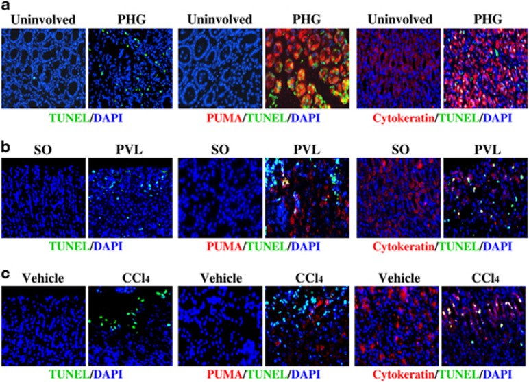
Figure 4.
PUMA-mediated apoptosis contributed to PHG. Apoptotic cells (green) were detected by TUNEL staining ( × 400), and PUMA (red) and cytokeratin (red) expression was determined by immunofluorescence staining ( × 400) in the gastric mucosal tissues. Cell nuclei (blue) were counterstained by DAPI ( × 400). (a) Gastric mucosal specimen of PHG patients. (b) Gastric mucosal specimen of PVL mice. (c) Gastric mucosal specimen of CCl4-treated mice

