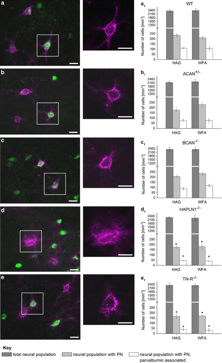
Figure 2.
Morphological and numerical appearance of neurons with and without PN in different mice knockout strains compared with wild type, detected in layers II and III of the barrel field cortex. Double fluorescence staining of PNs by WFA (pink) and the Ca-binding protein PV (green). Sections of wild-type mice (a) show intensely labelled, robust lattice-like structures enwrapping the neuronal soma and the proximal dendrites; PN of mice deficient for aggrecan (ACAN+/−) (b) or brevican (BCAN−/−) (c) look slightly fuzzy, but not disturbed. In contrast, PN of mice lacking the cartilage link protein (HAPLN1−/−) are attenuated and disturbed with almost no staining around dendrites. (d) Furthermore, Tenascin-R (TN-R−/−) knockout mice (e) display granular structures with lesser overall staining around dendrites. (a1–e1) Numerical density of neurons stained by NeuN (dark grey), of PN-associated neurons detected by (WFA, light grey) or an antibody against aggrecan core protein (HAG, light grey) and of PN-associated neurons expressing PV (white) in the barrel field cortex. In mice deficient for aggrecan (b1), link protein 1 (d1) or TN-R (e1), numbers of PN-associated as well as PN-associated, PV-expressing neurons were significantly reduced. The lack of brevican (c1) had no effect on the numerical density of neurons. Data are mean values, ±S.D. Statistical analysis by Mann–Whitney test, n=5. *P<0.05, scale bar 20 μm

