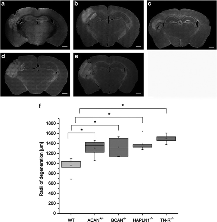
Figure 3.
Extent of neurodegeneration induced by single-sided injection of FeCl3 into the barrel field cortex of mice knockout strains lacking specific components of PNs. (a–e) Typical examples of coronal sections cut through the lesion at its maximal extension. A volume of 0.2 μl 20 mM FeCl3 was injected into the left barrel field cortex; degenerating cells were detected by Fluoro-Jade B staining 24 h later. Note that brain tissue volume affected by degeneration is significantly different in different mice strains. (a) WT; (b) ACAN+/−; (c) BCAN−/−; (d) HAPLN-1−/−, (e) TN-R−/−. Scale bar 500 μm. Quantification of lesion size determined on serial sections after staining with Fluoro-Jade B (f). In all mice knockout strains, lesion size was significantly larger than in wild type (WT). A lack of tenascin (TN-R−/−) results in the most extensive spreading of degeneration. In brevican knockout mice (BCAN−/−), heterozygous aggrecan knockout mice (ACAN+/−) and mice lacking the link protein 1(HAPLN-1−/−), lesion size is somewhat smaller. Data are expressed as mean values. Statistical analysis by Mann–Whitney test, n=5. *P<0.05

