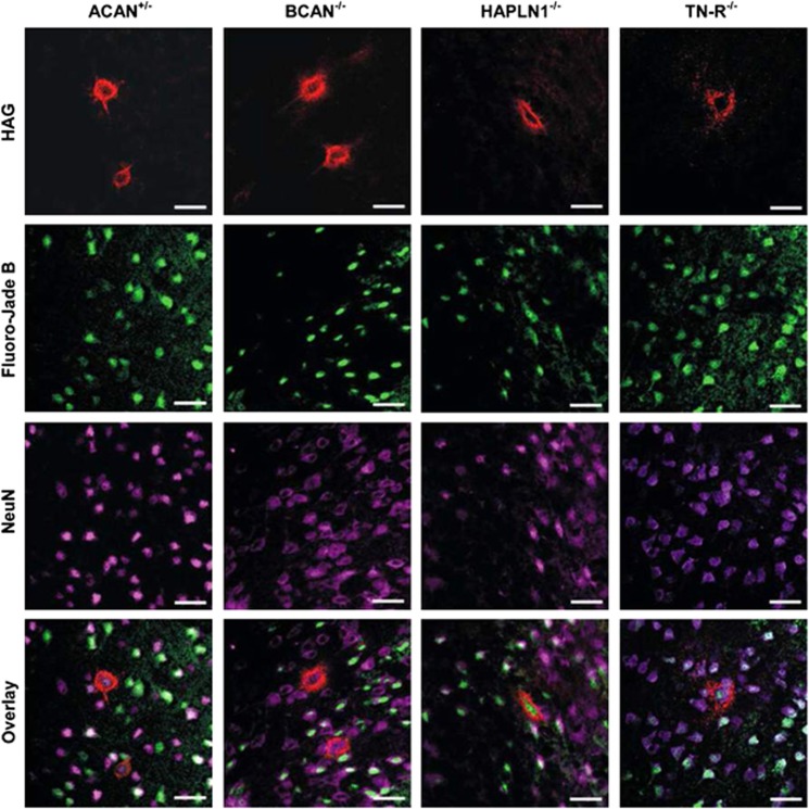
Figure 5.
Detection of death neurons with and without PN in different mice knockout strains detected by triple staining. PNs were visualized by HAG (red), degenerating cells were labelled by Fluoro-Jade B (green) and neurons were detected by NeuN (pink). Merged images were used for quantitative analysis. Scale bar 20 μm

