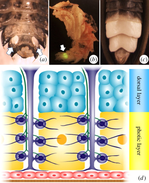
Figure 1.

(a) Ventral abdomen of a Photuris larva. Note the paired photic organs (POs) on the eighth abdominal segment (indicated by arrows). (b) Photuris pupa. The larval PO (indicated by arrow) remains functional throughout pupation and glows when the pupa is disturbed. (c) Close-up of the adult Photuris PO. (d) Diagrammatic cross section of adult photurid lantern. Note photic (yellow) and dorsal (blue) layers which are derived from fat body precursors [7]. Also shown are tracheae and tracheal end-cell complexes (purple), nerve axons (green), epidermis (red) and transparent cuticle (brown). Based on Chapman [8], after Smith [9].
