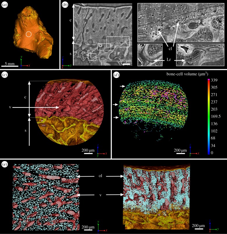
Figure 1.
Mid-shaft bone histology of the juvenile humerus of Eusthenopteron (NRM P246c). (a) Mesial view of the whole humerus showing the location of the high-resolution scan made at mid-shaft (voxel size: 0.678 μm). (b) Virtual thin section (made along the longitudinal axis) showing the primary bone deposit of cortical bone and its connection to the spongiosa. Some remnants of calcified cartilage are still preserved at the location of Katschenko's line (chondrocyte lacunae) and within the spongiosa (Liesegang rings). (c) Transverse view of the three-dimensional organization of the vascular mesh embedded within the cortical bone and the underlying trabecular spongiosa. (d) Quantification of the volume of bone cells showing three recurrent periods of volume decrease (green layers pointed out with white arrows) interpreted as phases of decreased growth. (e) From left to right: top and longitudinal views of the vascular mesh showing the circular and radial alignment of the vascular canals (in pink). The osteocyte lacunae are represented in bright blue. c, cortical bone; cl, chondrocyte lacunae; Lr, Liesegang rings; ol, osteocyte lacunae; s, spongiosa; v, vascular mesh.

