Figure 2.
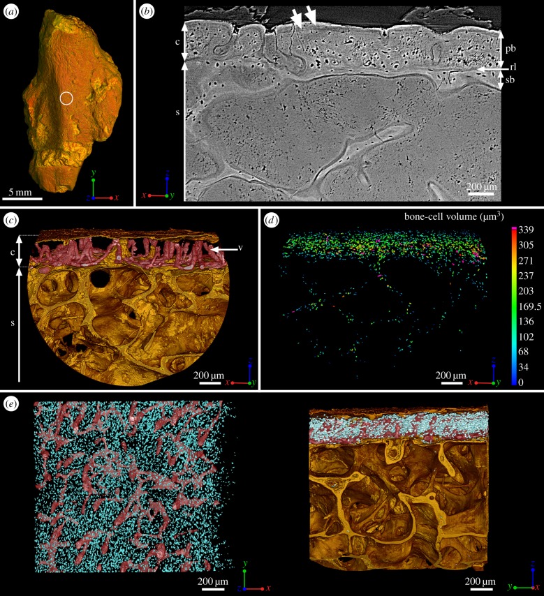
Mid-shaft bone histology of the adult humerus of Eusthenopteron (NRM P248d). (a) Mesial view of the whole humerus showing the location of the high-resolution scan made at mid-shaft (voxel size: 0.678 μm). (b) Transverse virtual thin section showing the primary bone deposit of cortical bone, the innermost part of which has been drastically eroded. Although X-ray tomography does not allow the nature of the bone matrix (i.e. the collagen fibre organization) to be determined, secondary bone can be distinguished from primary cortical bone because it is always demarcated by a resorption line. A secondary bone deposit of cellular endosteal bone is laid down on the inner surface of the primary cortex. The trabeculae of the spongiosa are also covered by a thin layer of cellular endosteal bone. (c) Transverse view of the three-dimensional organization of the vascular mesh embedded within the cortical bone and the underlying trabecular spongiosa. (d) Quantification of the volume of bone cells showing an obvious decrease of volume just under the surface of the primary bone and in the endosteal bone (blue) (cf. electronic supplementary material, figure S8 for detail). (e) From left to right: top and longitudinal views of the vascular mesh showing a non-oriented organization of the vascular canals (same colour code as for figure 1). c, cortical bone; pb, primary bone; rl, resorption line; s, spongiosa; sb, secondary bone; v, vascular mesh.

