Figure 7.
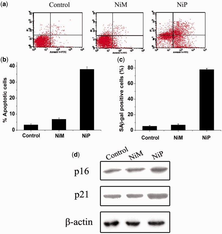
Apoptosis and senescence evoked by NiP-induced telomere dysfunction. (a) Apoptotic cell death induced by NiP in MCF-7 cells. After treatment with NiP, cells were collected and stained with PI and Annexin V-FITC. Representative data of flow cytometry was shown. (b) Annexin V positive/PI negative cells were measured by flow cytometry. The percentages of cells undergoing apoptosis were expressed with respect to the total number of cells. (c) Expression of senescence-associated β-galactosidase (SA-β-gal) in MCF-7 cells after continuous treatment with NiM or NiP. This assay was performed in triplicate. The senescent cells were counted under an inverted microscope in five random fields. (d) Upregulation of p16 and p21 proteins induced by NiP. After MCF-7 cells were treated with NiM or NiP, cells were lysed and separated on 10% SDS-PAGE and probed with anti-p16INK4a and anti-p21WAF1 primary antibody, respectively. Immunoblotting for β-actin was also performed to verify equivalent protein loading. Each experiment has been repeated three times.

