Figure 4.
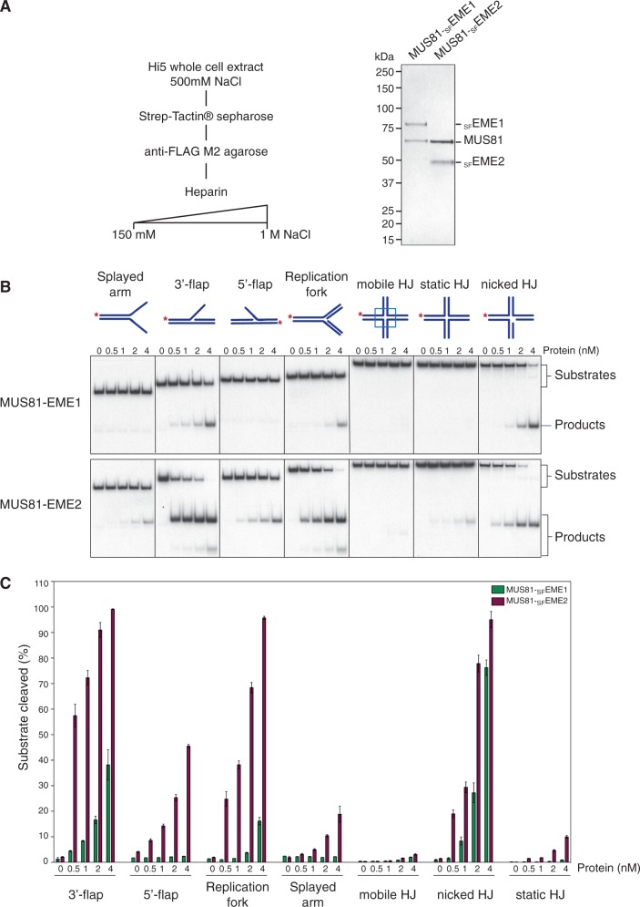
Substrate specificities of human MUS81-SFEME1 and MUS81-SFEME2. (A). Purification scheme and visualization of purified human MUS81-SFEME1 and MUS81-SFEME2 by SDS–PAGE followed by staining with InstantBlue. (B). The 32P-labelled DNA substrates (100 nM) were incubated with the indicated concentrations of purified MUS81-EME1 or MUS81-EME2 for 30 min at 37°C, and the products were analysed by neutral PAGE and visualized by autoradiography. The 5′-32P-end labels are indicated with asterisks. (C). Quantification of the data shown in Figure 4B was performed by phosphorimaging analysis. Product formation is expressed as a percentage of total radiolabelled DNA. Data are presented as the mean of three experiments (±SEM).

