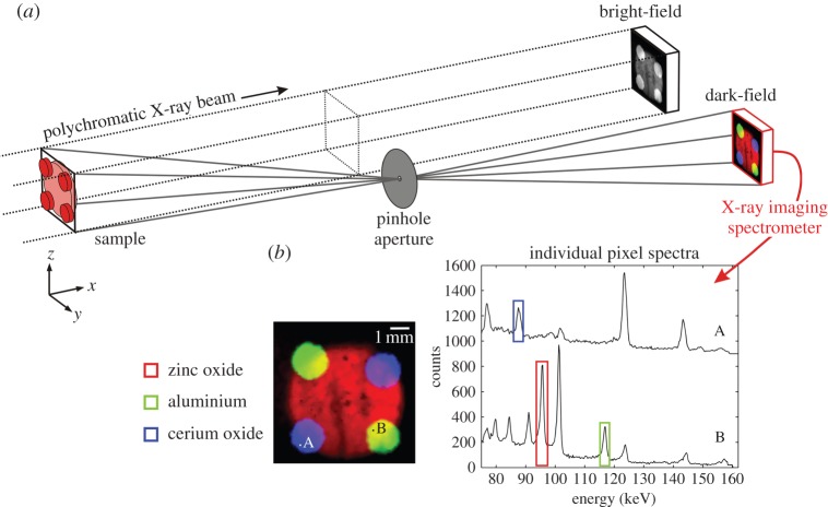
Figure 1.
Dark-field hyperspectral X-ray imaging. (a) A flood-field polychromatic X-ray beam illuminates a sample, which after attenuation yields a bright-field image. The dark-field is imaged using an off-axis pinhole aperture projecting signals onto an X-ray imaging spectrometer. (b) Individual pixel spectra show distinct diffraction peaks corresponding to different materials within the sample. By integrating over specific spectral bands materials can be mapped accordingly.

