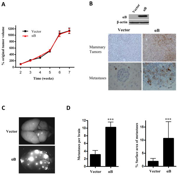
Figure 5. αB-crystallin overexpression increases brain metastases in an orthotopic TNBC model.
231-mCherry cells stably expressing vector or αB-crystallin (αB) were injected bilaterally into the ducts of the 4th mammary gland of NSG mice. (A) Mammary tumor volume expressed as the percentage original tumor volume at 2 weeks in mice with 231-mCherry-Vector and 231-mCherry-αB xenografts (n=10 mice per group). (B) Immunoblot of 231-mCherry-Vector and 231-mCherry-αB mammary tumors and αB-crystallin IHC staining of mammary tumors and brain metastases in both groups. (C) Representative fluorescent whole brain images from mice with 231-mCherry xenografts overexpressing vector or αB. (D) Number of mCherry-fluorescent metastases per brain in vector and αB groups, and percentage surface area of brain metastases in vector and αB groups (mean ± SEM, n = 10 mice per group, ***P < 0.001).

