
Figure 1.
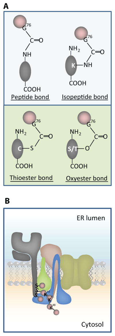
A. Schema c showing different chemical bonds between Ub and substrates. The upper panel shows pep de and isopep de linkages between Ub (Pink balls) and protein substrates (Gray ovals). Ub coupling on the N-terminus is shown on the le# and Ub coupling to an internal K residue is shown on the right. The lower panel of A shows thio- and oxy-ester linkages of Ub to C or S/T residues of substrates, respec vely. B. Schema c model showing how viral ligase mK3 (blue) interacts with Ube2j2 (green) to ubiqui nate S residues on the cytosolic tail of MHCI heavy chain (HC) (gray), then build a Lys48 (K48) linked chain. The adaptor proteins TAP/tapasin that confer substrate specificity by orien ng mK3/Ube2j2 with the HC tail are shown in yellow/tan.