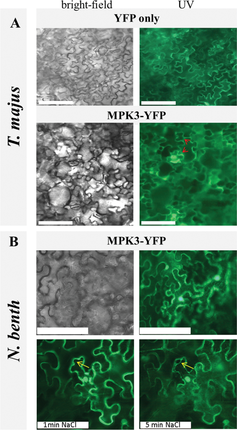
Figure 4.
Subcellular Localization of MPK3 In Planta.
Transient expression of free YFP (top) or MPK3–YFP (bottom) in agro-infiltrated T. majus (A). Regions of putative MPK3 membrane associations are indicated by arrows.
(B) Transient expression of MPK3–YFP and plasmolysis studies in N. benthamiana. MPK3–YFP localization was documented by UV microscopy 4 d post infiltration directly (top, right) and 1 or 5min after treatment of leaf discs with a 2-M NaCl solution (bottom). The progressive detachment of the plasma membrane in NaCl-treated cells is marked by an arrow. Scale bar: 100 μm.

