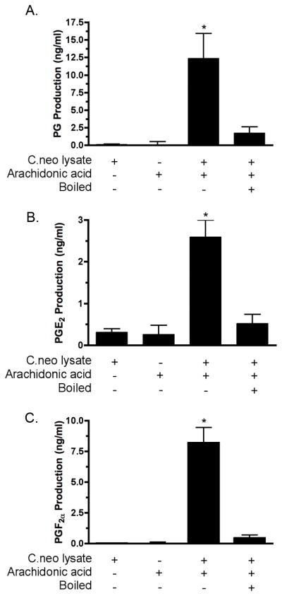
Figure 1. Prostaglandin production in cryptococcal lysates.

C. neoformans cells were lysed and the lysates incubated ± AA for 2 hr. at 37°C. C. neoformans lysates were incubated with AA and total prostaglandins were measured using a prostaglandin screening EIA (Panel A (n = 11; * = p < 0.001)); PGE2 were levels assayed using a PGE2 monoclonal EIA (Panel B (n = 7; * = p < 0.001)); and PGF2α levels were assayed using a PGF2α monoclonal EIA (Panel C (n = 5; * = p < 0.001)).
