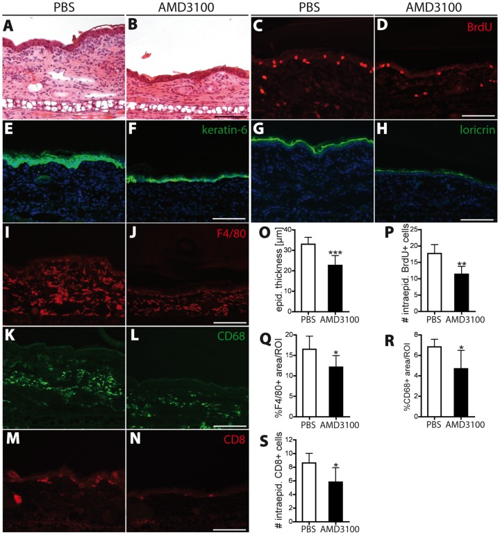
Figure 5. Inhibition of CXCR4 reduces inflammatory cell infiltration into the skin and normalizes epidermal architecture.
(A–B) H&E stains of ear skin sections at day 21 showed that AMD3100 treatment reduced edema formation, epidermal thickening and inflammatory cell infiltration. (C–D) CXCR4 inhibition reduced the number of intraepidermal BrdU+ proliferating cells in the inflamed ear skin. (E–H) The hyperproliferation-associated keratin 6 and loricrin, a marker of terminal epidermal differentiation, were less broadly expressed in the epidermis of AMD3100-treated mice than in PBS-treated mice. (I–L) Immunofluorescence staining of the two macrophage markers F4/80 and CD68 revealed a significant reduction in the percentage of area covered by macrophages in AMD3100-treated mice compared to PBS treatment. (M–N) Inhibition of CXCR4 decreased the number of intraepidermal CD8+ T-cells in the inflamed ear skin. One ear half is shown. (O–P) Computer-assesed quantification of epidermal thickness (O), number of intraepidermal BrdU+ cells (P), the percentage of covered area by F4/80 (Q) and CD68 (R) postitive macrophages and the number of intraepidermal CD8+ cells (S). Scale bars represent 100 μm. Data represent mean ±SD. *P<0.05; **P<0.01; ***P<0.001.

