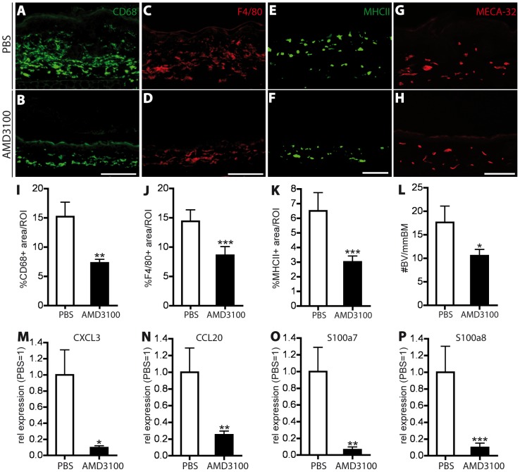
Figure 7. CXCR4 inhibition reduces inflammatory cell infiltration, angiogenesis and inflammatory marker expression in the imiquimod-induced skin inflammation.
(A–H) Representative images of immunofluorescence stains for CD68+ (A, B) and F4/80+ (C, D) macrophages, MHCII+ antigen presenting cells (E, F) and MECA-32+ blood vessels (G, H) in the IMQ-inflamed ear skin of PBS and AMD3100-treated mice. One ear half is shown. Scale bars represent 100 μm. (I–K) Quantitative image analysis showed a significant reduction in the percentage of area covered by macrophages (I, J) and antigen presenting cells (K) in AMD3100-treated mice. (L) Inhibition of CXCR4 decreased the number of Meca-32+ blood vessels (BV). (M–P) Real-time RT-PCR analyses of RNA from whole ear skin extracts of imiquimod-inflamed mice showed a significant downregulation of CXCL3 (M), CCL20 (N), S100a7 (O) and S100a8 (P). Data represent mean ±SD. *P<0.05; **P<0.01; ***P<0.001.

