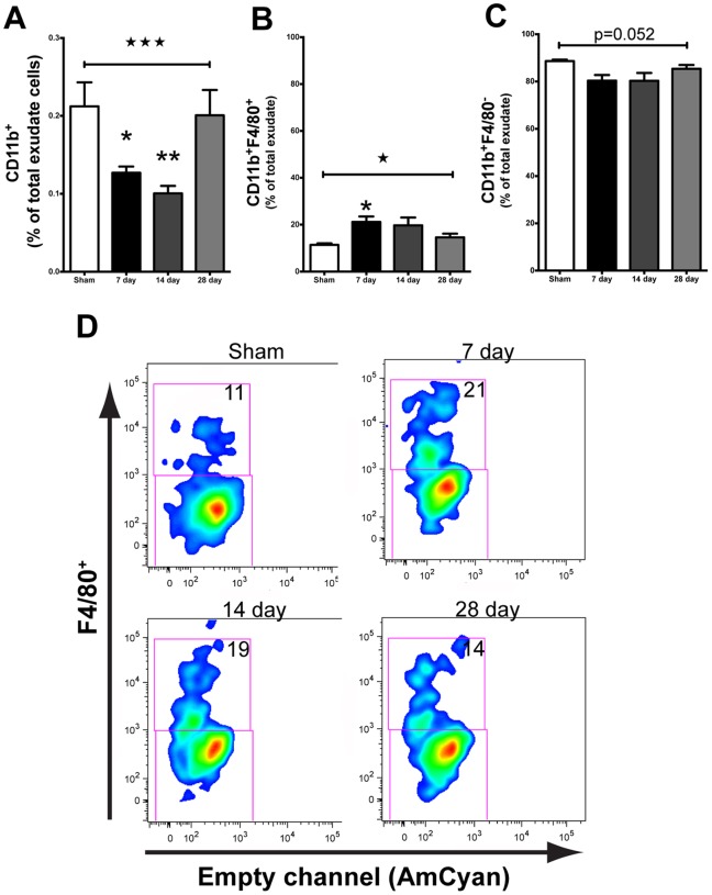
Figure 1. Cranial irradiation alters myeloid cell population over time.
A. 10+ cells (ANOVA; F(3,12) = 12.07, ★★★p = 0.0006) at 7 (Tukey’s HSD; *p<0.05) and 14 (Tukey’s HSD; **p<0.01) days post irradiation. The percentage of CD11b+ cells returned to sham levels by day 28 (Tukey’s HSD; p>0.05). B. Overall, cranial irradiation induced a significant increase in the percentage of CD11b+F4/80+ macrophages at 7 days (ANOVA; F(3,12) = 4.048, ★p = 0.0335; Tukey’s HSD; *p<0.05). C. Concomitant with the radiation induced increase of percentage of F4/80+ macrophages, we observed a trend for decreased F4/80− stained cells. D. Representative plots showing the 10 Gy radiation induced shift in the percentage of CD11b+ cells that stained for F4/80 as a function of time. Average proportion of F4/80+ is given.

