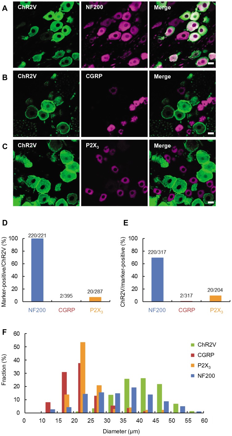
Figure 1. Distribution of channelrhodopsin 2-Venus conjugates (ChR2V) in the trigeminal ganglion (TG) neurons.
A-C, Immunohistochemical identification of ChR2V-positive (ChR2V+) neurons using cell-type specific markers, NF200 (A), CGRP (B) and P2X3 (C). Scale bars indicate 20 μm. D, Probability of the co-expression of each marker, NF200, CGRP or P2X3, in the ChR2V+ neurons. E, Probability of the co-expression of ChR2V in the neurons positive for each marker, NF200, CGRP or P2X3. F, The average diameters of the TG neurons were compared among four groups positive for ChR2V (green), NF200 (blue), CGRP (red) and P2X3 (orange).

