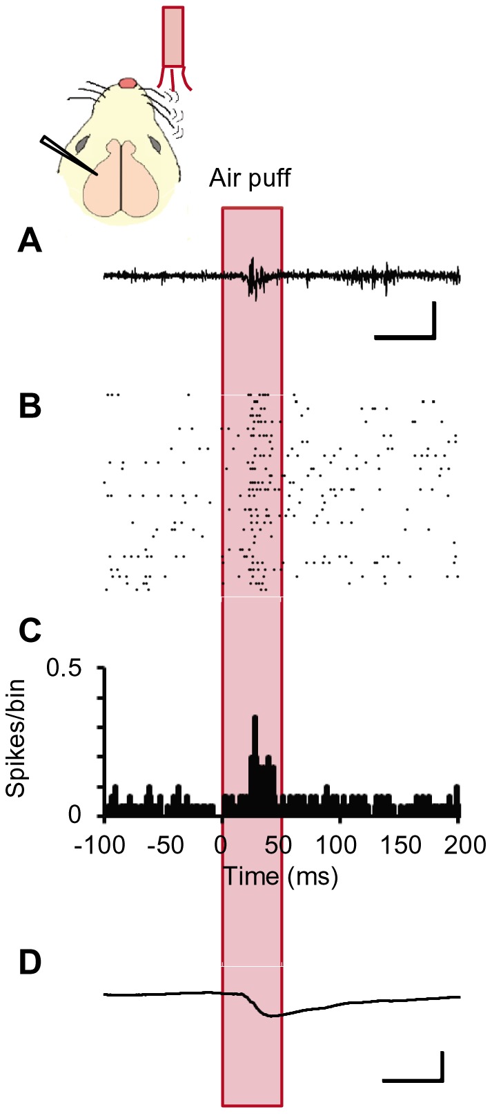
Figure 4. Barrel cortical activities evoked by whisker mechano-stimulation.

A-D, A sample MUA recording trace (A), raster plots of spikes (B), peristimulus-time histograms (PSTHs, bin width = 1 ms) of spikes (C) and the averaged LFP record (D) in response to the air puff (duration, 50 ms) to the contralateral whiskers. Scales, 50 ms and 100 μV (A) or 0.5 mV (D).
