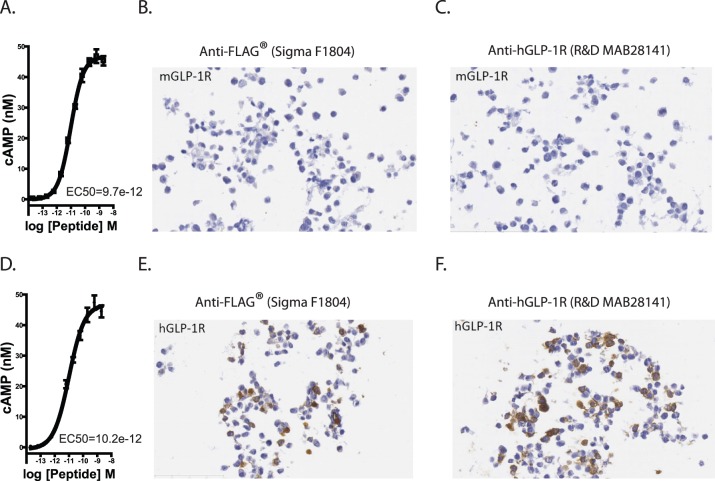
Figure 5. Validation of GLP-1R and FLAG antibodies.
(A) mGlp-1r and (D) hGLP-1R were transiently expressed in HEK cells. cAMP accumulation in response to GLP-1 showed similar cAMP accumulation. HEK cells expressing mGLP-1R showed no (B) FLAG or (C) FLAG-tagged hGLP-1R expressing. HEK cells show stained for (E) FLAG and (F) hGLP-1R protein after transient transfection.

