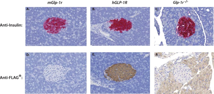
Figure 6. IHC of pancreata from mGlp-1r, hGLP-1R, and Glp-1r−/− mice.
Pancreata were stained for insulin and showed positive staining in the β-cells of (A) mGlp-1r, (B) hGLP-1R, and (C) Glp-1r−/− islets. FLAG staining was also performed and (E) hGLP-1R islets stained positive for FLAG with none detected in (D) mGlp-1r and (F) Glp-1r−/− islets.

