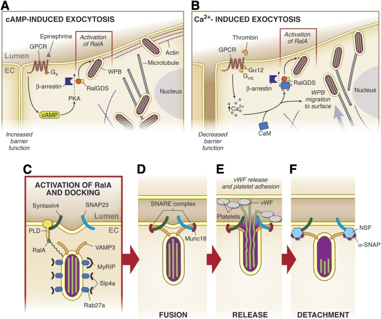
Fig. 9.
The exocytosis microdomain. (A) Epinephrine induces increased cAMP via Gs protein-coupled receptors, resulting in the activation of RalA by RalGDS. The exocytosis of the Weibel-Palade bodies present at the PM is activated, whereas the progression of the WPBs located in the perinuclear region is inhibited by the presence of a more prominent peripheral actin rim. This process is accompanied by an increase in barrier function. (B) Upon activation of GqPCR in ECs by agonists such as histamine or thrombin, there is an increase in [Ca2+]i, which associates with CaM. The Ca2+-CaM further binds to the amino terminus of Ral GDS, which leads to the dissociation from the inhibitory beta-arrestin and to activation of RalA. This whole process allows WPB migration to the surface and concurrently decreases the strength of the barrier function between ECs. (C) RalA promotes fusion of the membrane by increasing PLD activity. Rab27a helps to determine when exocytosis will occur via its ratio of fractional occupancy by MyRIP and Slp4a. (D) The V-SNARE VAMP3 and the t-SNAREs syntaxin4 and SNAP23 interact to pull the two membranes in close proximity for fusion to occur. Munc18 acts to inhibit the SNAREs from binding prematurely. (E) vWF is released into the lumen, where it can bind to and attract platelets in addition to exerting effects on neighboring ECs. (F) NSF/α-SNAP bind to SNAREs to facilitate their disassembly.

