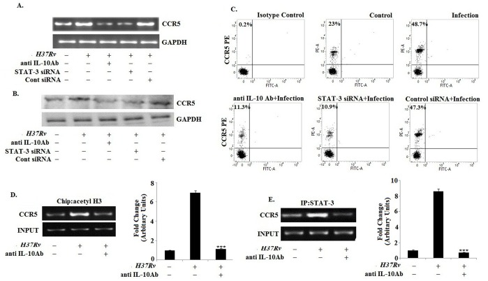
Figure 4. IL-10 augments the CCR5 expression in H37Rv infected macrophages via involving STAT3.
Bone marrow derived macrophages (2×10⁶cells/ml) were pretreated with either anti IL-10 Ab (10 ug/ml) or with control siRNA and STAT3-specific siRNA and then infected with Mycobacterium tuberculosis H37Rv (MOI = 1∶10). Changes in messenger RNA (mRNA) expression of CCR5 and GAPDH were determined by semi quantitative RT-PCR (A). In a separate set, the pretreated and infected macrophages were lysed and subjected to Western blot with anti-CCR5 antibody as described in Materials and Methods (B). Infected macrophages were analyzed by flow cytometry for CCR5 (PE) expression as described in figure legend 1 (C). Data represented here are from one of three independent experiments, all of which yielded similar results. Murine macrophages (1×10⁶cells/ml) were treated with anti IL-10 Ab for 1 h and then subsequently followed by Mycobacterium tuberculosis infection for 45 min. After 45 min of incubation, ChIP assays were conducted as described in Materials and Methods. Immunoprecipitations were performed using Abs specific to acetylated H3 (IP acetyl-H3) (D) or STAT-3 (E), and conventional RT-PCR was performed using primers specific to the CCR5 promoter. Data represented here are from one of three independent experiments, all of which yielded similar results.

