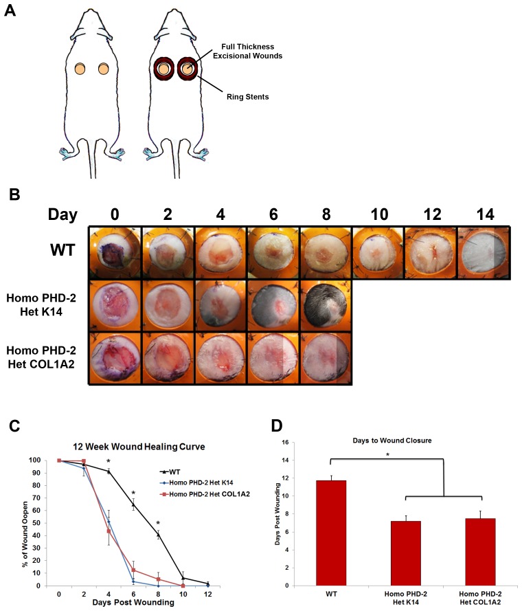
Figure 2. In vivo data from humanized wound healing model.
A) Schematic of stented full-thickness excisional wound model. B) Representative pictures of wounds from days 0–14 for wild type, heterozygous K14-Cre/homozygous floxed PHD-2, and heterozygous Col1α2-Cre-ER/homozygous floxed PHD-2 mice. C) Twelve week wound healing curve showing the wound closure rate for wild type, heterozygous K14-Cre/homozygous floxed PHD-2, and heterozygous Col1α2-Cre-ER/homozygous floxed PHD-2 mice with significant differences on days 4, 6, and 8 (*p<0.05). D) Graph representing days to wound closure with a significant difference in days to closure between the wild type and the heterozygous K14-Cre/homozygous floxed PHD-2 and heterozygous Col1α2-Cre-ER/homozygous floxed PHD-2 mice (*p<0.05).

