Figure 5.
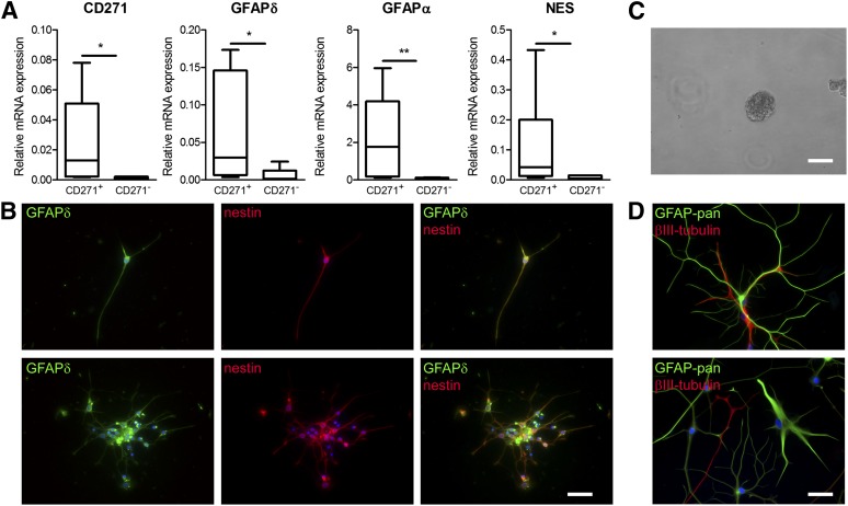
Isolation of CD271+/GFAPδ+ neural progenitor cells derived from the subventricular zone of patients with a neurodegenerative disorder. (A): mRNA expression levels in CD271+ and CD271− fractions directly after isolation. Data are expressed as median and interquartile ranges. Analysis of significance was performed by Mann-Whitney U test. n = 6 donors, *p < .05, **p < .01. (B): Immunofluorescent double labeling of CD271+ cells 5 days after isolation. (C): CD271+/GFAPδ+ cells derived from a donor with Alzheimer’s disease (AD)-formed neurospheres in vitro. (D): CD271+/GFAPδ+ cells derived from an AD donor were able to differentiate into GFAP-positive astrocytes (green) and βIII-tubulin-positive neurons (red). Scale bar represents 250 μm (C) and 50 μm (B, D). Abbreviations: GFAP, glial fibrillary acidic protein; NES, nestin.

