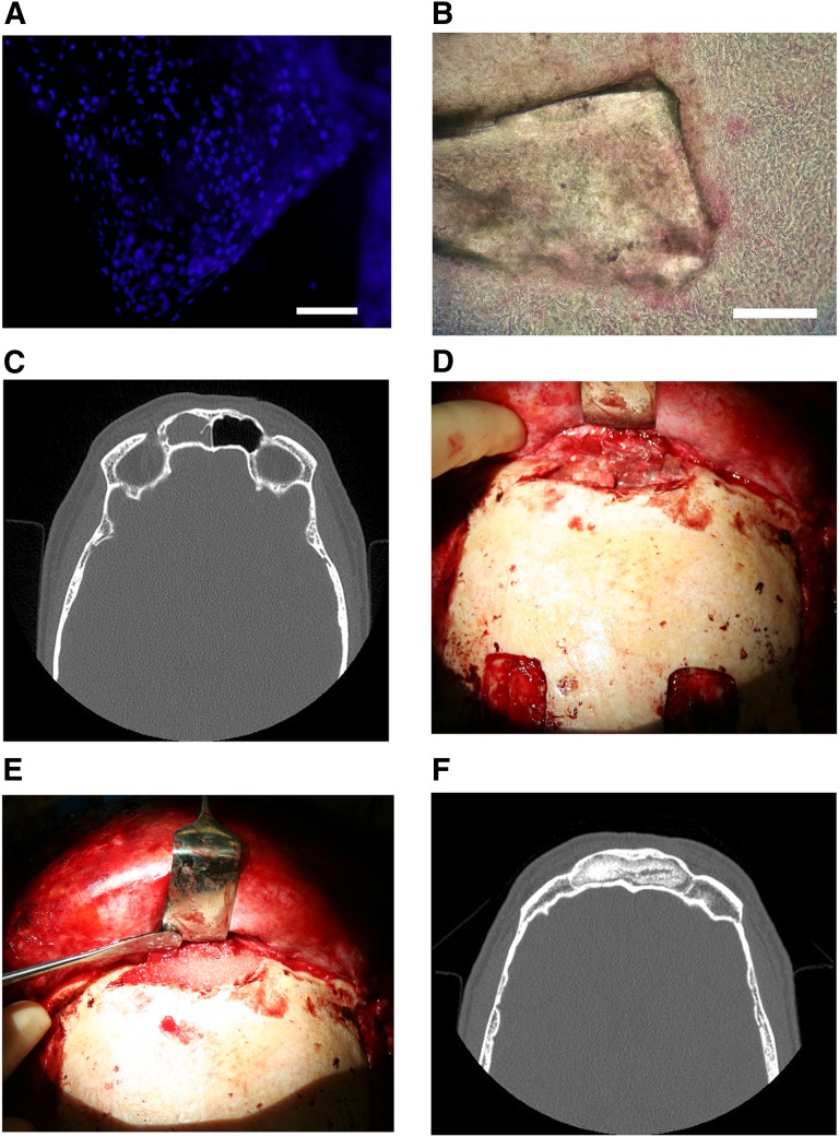
Figure 1.
Collage of frontal sinus treatment. (A): 4′,6-diamidino-2-phenylindole nuclear staining image of bioactive glass granules seeded with adipose stem cells (BonAlive). Scale bar = 1 mm. (B): Alkaline phosphatase staining of a bioactive glass granule seeded with adipose stem cells (BonAlive). Scale bar = 1 mm. (C): Preoperative computed tomography (CT) scan of the left frontal sinus demonstrating mucosal changes and frontal sinusitis. (D): Clinical photograph of exposed diseased frontal sinus with purulent mucosa. (E): Clinical photograph of debrided frontal sinus packed with granules of bioactive glass seeded with autologous adipose-derived stem cells. (F): Postoperative CT scan of the frontal sinus obliterated with autologous adipose stem cell-seeded bioactive glass 28 months following surgery with no resorption of the construct.

