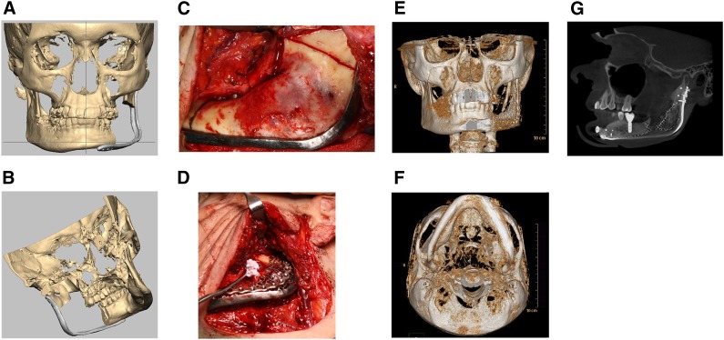
Figure 3.
Collage of mandibular reconstructions. (A): Virtual preoperative planning using Romexis software with computer-generated image of patient with large mandibular ameloblastoma showing the reconstruction plate over the area planned for resection from an anterior view. (B): Computer-generated image of patient with large mandibular ameloblastoma showing the reconstruction plate over the area planned for resection from a medial view. (C): Intraoperative photograph showing reconstruction plate in position and resection lines on mandibular ramus posteriorly and body anteriorly. (D): Intraoperative photograph with adipose-derived stem cell-seeded β-tricalcium phosphate (β-TCP) granular construct with recombinant human bone morphogenetic protein-2 (rhBMP-2) being placed beneath titanium containment mesh at mandibular resection site. (E): Postoperative three-dimensional (3D) computed tomography (CT) scan anterior view of the regenerated left body and ramus of the mandible 12 months after reconstruction. (F): Postoperative 3D CT scan basilar view of the regenerated left body and ramus of the mandible 12 months after reconstruction. (G): Cone beam CT scan showing mandibular reconstruction using autologous adipose-derived stem cell-seeded β-TCP granular construct with rhBMP-2 restored with successful functionally loaded dental implant in the regenerated bone.

