
Figure 2. TGF-β is necessary and sufficient for neuronal C1q upregulation in vitro.
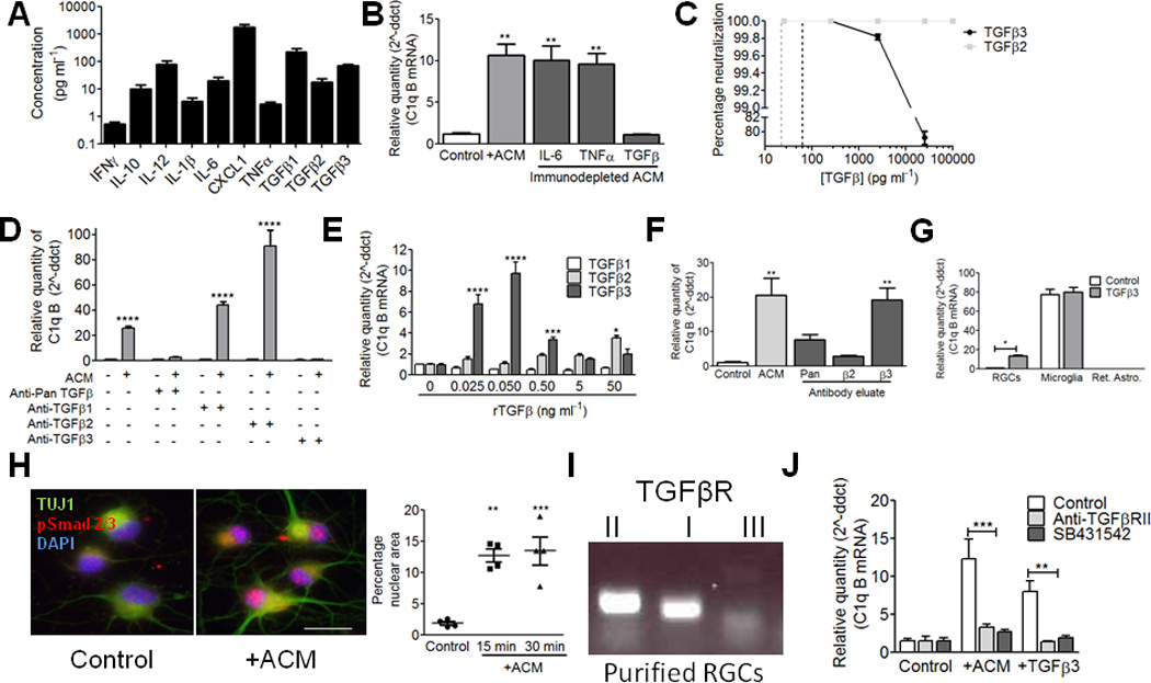
(A) ACM cytokine profiling by ELISA. N=3 independent ACM batches. (B) Immunodepletion of TGF-β, but not other cytokines, significantly reduced ACM-induced C1q upregulation (one-way ANOVA, n= 3 experiments, ***p<0.001, **p<0.01, F(4,12)=22.15). (C) Validation of immunodepletion. %Neutralization represents ([TGF-β]initial – [TGF-β]depletion)/(initial TGF-β concentration). N=3 experiments. (D) Immunodepletion of each TGF-β isoform showed that depletion of pan-TGF-β or TGF-β3 blocked C1q upregulation (two-way ANOVA, n=3 experiments, ***p<0.001, F(4,20)=79.52). (E) Concentration-response curves for TGF-β1, 2, and 3 (Two-way ANOVA, n= 3 experiments, ****p<0.0001, **p<0.01, F(10,36)=23.56). (F) Glycine elution showed that either anti-pan TGF-β or anti-TGF-β3 eluates upregulate C1q (one-way ANOVA, n= 3 experiments, ***p<0.001, F(4,10)=11.02). (G) QPCR results for c1qb in RGC cultures and in microglia or retinal astrocyte cultures after TGF-β3 treatment (50 pg/ml) for 15 min. (two way ANOVA, n=3 experiments, p<0.05, F(2,12)=415.96). (H) RGCs showed increased nuclear accumulation of pSmad2 (15–30 min. ACM treatment). Quantification showed a significant increase in pSmad (red) within the nuclear area (blue) (one-way ANOVA, n= 15 cells/condition, ***p<0.001, F(2,9)=19.83). Scale bar = 20um. (I) RT-PCR for tgfbr1, tgfbr2, and tgfbr3 in P5 retina. Data shown are representative of 4 samples tested. Full length gels are displayed in Supplemental Fig. 6. (J) Blocking TGFβRII signaling with neutralizing antibodies or with inhibitors of TGFβRI significantly reduced the effects of ACM or TGF-β3 (0.05 ng/ml) on C1q (two-way ANOVA, n= 3 experiments, **p<0.01, ***p<0.001, F(4,18)=35.06).