Figure 2.
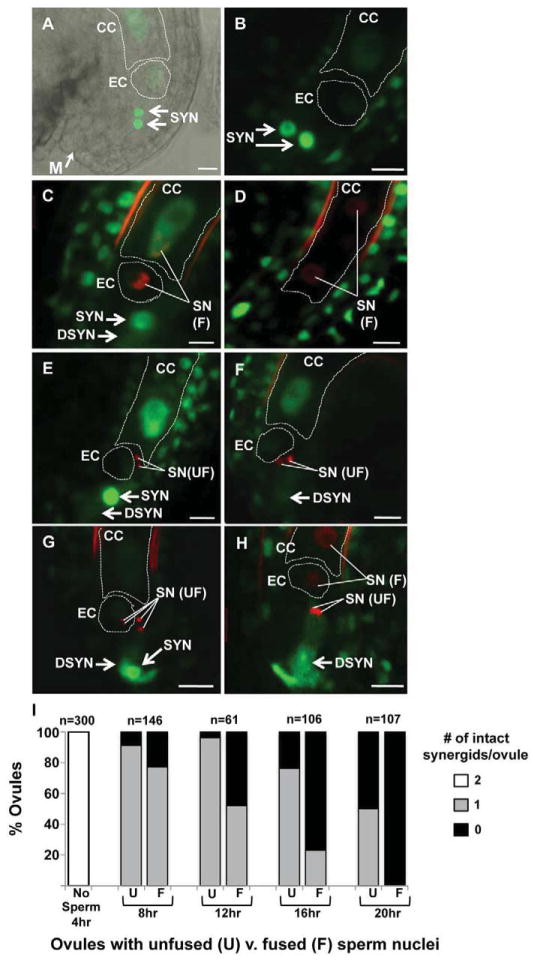
The remaining synergid cell persists longer in ovules targeted by nonfunctional sperm. (A) An unfertilized ovule expressing ACT11:MSI1:GFP. DIC and confocal images are overlaid to highlight the position of the micropyle (M, same in all panels) and GFP accumulation in the egg (EC), central (CC) and synergid (SYN) nuclei. (B–H) Pistils were pollinated with hap2-1/HAP2(GCS1), HTR10:HTR10:mRFP. Representative confocal micrographs are shown of an unfertilized ovule (B), ovules that have received sperm and have a persistent synergid (SYN) (C,E,G), or ovules that have received sperm and both synergids have degenerated (DSYN) (D,F,H). (D) ACT11:MSI1:GFP signal was not detectable in synergids because this image was obtained later in development (note dividing primary endosperm) and significantly after synergid degeneration. (I) Ovules were scored for the number of synergids that remain intact (2, white bar; 1 gray bar; 0 black bar) and whether they had received sperm whose nuclei fused with female nuclei (fused, F) or sperm that remained unfused (U). Data are reported as the percentage of the total number of ovules analyzed (n) at each time point following pollination (hours, hr). Scale bar, 10μm.

