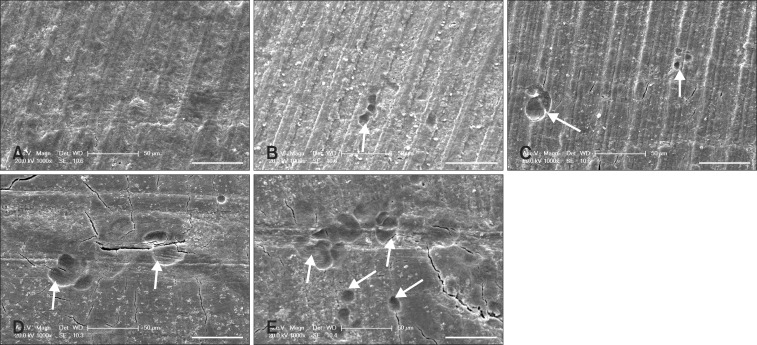
Fig. 4.
Resorption lacunae on bone slices observed with an XL30-ESEM environmental scanning electron microscope. No resorption lacunae were observed on bone slices incubated with cells from group A (RAW264.7 cells cultured without any cytokines). In contrast, many resorption lacunae (indicated with white arrows) were formed in bone slices incubated with cells from groups B ~ E. (A) No cytokines. (B) 30 µg/L RANKL and 25 µg/L M-CSF. (C) 30 µg/L RANKL, 25 µg/L M-CSF, and 10-10 mol/L 1α, 25-(OH)2D3. (D) 30 µg/L RANKL, 25 µg/L M-CSF, and 10-9 mol/L 1α, 25-(OH)2D3. (E) 30 µg/L RANKL, 25 µg/L M-CSF, and 10-8 mol/L 1α, 25-(OH)2D3. Scale bars = 50 µm.

