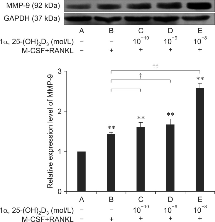
Fig. 6.
Detection of MMP-9 protein expression in osteoclasts by Western blotting. The results are expressed as the mean ± SE. MMP-9 was expressed in each group of cells. Compared to group A (RAW264.7 cells cultured without any cytokines), RANKL and M-CSF enhanced the expression of MMP-9 (**p < 0.01 vs. control group). Furthermore, 1α,25-(OH)2D3 promoted the expression of MMP-9 in a dose-dependent manner (†p < 0.05 vs. group B and ††p < 0.01 vs. group B).

