Fig. 2.

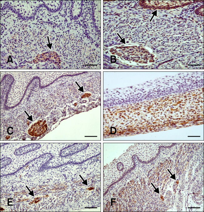
Distribution of immunoreactivity in the goat rumen. (A) Synaptophysin (SPY)-positive staining in the tunica muscularis and myenteric plexus (arrow) in a section of rumen wall at 64 days (43% gestation). (B) Non-neuronal enolase (NNE) immunoreactivity in the epithelial layer (arrow), lamina propria-submucosa, tunica muscularis, and myenteric plexus (arrow) at 64 days (43% gestation). (C) Glial fibrillary acidic protein (GFAP)-positive staining at 64 days (43% gestation) in the lamina propria-submucosa, tunica muscularis, serosa, and myenteric plexus, (arrows) and within ruminal papillae. (D) Vimentin (VIM)-specific immunostaining in the mesenchymal cells and serosa of the ruminal wall at 39 days (25% gestation). (E) Neuropeptide Y (NPY)-positive staining in the lamina propria-submucosa, tunica muscularis, and myenteric plexus (arrows) at 113 days (75% gestation). (F) Vasoactive intestinal peptide (VIP)-specific staining at 113 days (75% gestation) in the lamina propria-submucosa. Intense immunostaining was observed in the tunica muscularis and myenteric plexus (arrows). Scale bars = 20 µm (D), 25 µm (A, B, E, and F), or 30 µm (C).
