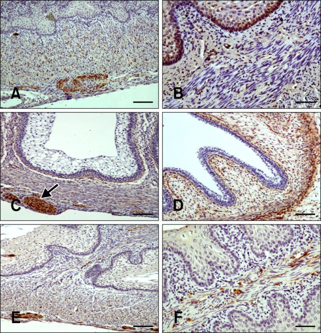
Fig. 4.

Distribution of immunoreactivity in the goat omasum. (A) SYN-positive staining in the lamina propria-submucosa, tunica muscularis, and myenteric plexus in the omasal wall at 75 days (50% gestation). (B) NNE-specific immunoreactivity in the epithelial layer, lamina propria-submucosa, and tunica muscularis at 64 days (75% gestation). (C) GFAP-positive staining within the myenteric plexus (arrow), lamina propria-submucosa, tunica muscularis, and serosa at 69 days (46% gestation). (D) VIM-specific immunostaining in the pluripotential blastemic tissue layer and serosa at 39 days (25% gestation). (E) NPY-positive staining in the lamina propria-submucosa, tunica muscularis, and myenteric plexus. An immunostained surface was visible in the connective tissue of the omasal laminae at 95 days (63% gestation). (F) VIP-positive staining in the connective tissue and tunica muscularis of the omasal laminae at 113 days (75% gestation). Scale bar = 25 µm (A, C, and E) or 20 µm (B, D, and F).
