
Fig. 3.
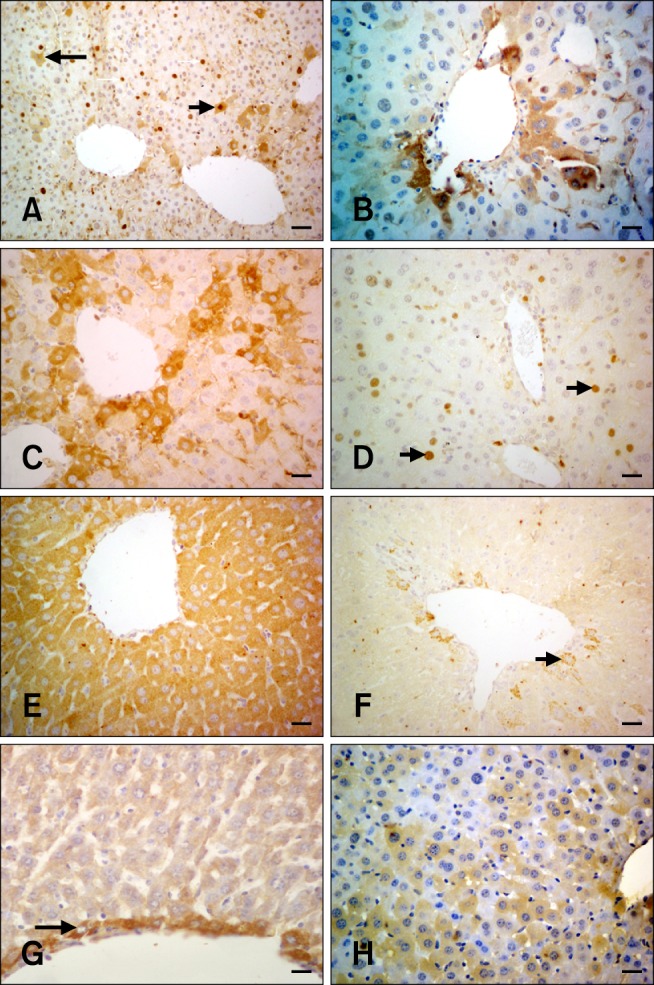
PCNA, TNFα, and FGF-2 immunohistochemistry in hepatocytes from the experimental and control groups. (A) Only the nuclei are positive for PCNA (white arrows). Only cytoplasmic PCNA staining was observed in some hepatocytes while the nucleus was negative (long black arrow). Both cytoplasmic and nuclear staining with the PCNA antibody (short black arrow; Group 6B) is shown. (B) Cytoplasmic PCNA-specific staining in periacinar hepatocytes (Group 4B). (C) Intense cytoplasmic PCNA staining of the periacinar hepatocytes on the 14th day (Group 4A). (D) Decreased PCNA expression in the hepatocytes (arrows; Group 5A). (E) Diffuse cytoplasmic staining specific for TNFα; the nucleus is negative (Group 3C). (F) Cytoplasmic TNFα-specific staining of some periacinar hepatocytes (arrow; Group 5C). (G) Hepatocytes surrounding the central vein were positive for FGF-2 (arrow; Group 1A). (H) Cytoplasmic FGF-2 expression (Group 4A). Scale bars = 18 µm (G), 22 µm (B ~ F and H), and 44 µm (A).