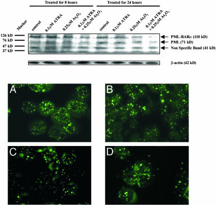
Fig. 2.
(Upper) Western blot analysis of PML-RARα fusion protein in cultured NB4 cells. Note that the intensity of the band corresponding to PML-RARα (arrow, 110 kDa) decreased on cotreatment with ATRA/As2O3 within 8 h. After 24 h of incubation, this band was almost undetectable in the eighth lane, and obvious degradation was observed in cells treated with 0.25 μM As2O3. (Lower) Immunofluorescence analysis of the subcellular localization of PML-RARa/PML in cultured fresh APL cells(original magnification ×1,000). (A) Untreated; (B) 8 h after 0.1 μM ATRA treatment; (C) 8 h after 0.25 μM As2O3 treatment; (D) 8 h after cotreatment with ATRA/As2O3 at the same concentrations.

