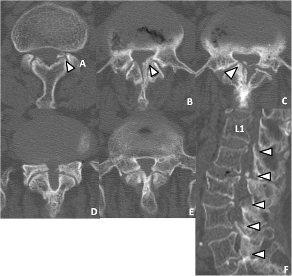
Figure 1.

Preoperative computed tomography. (A) through (E) Preoperative computed tomographic scans show congenital spinal stenosis from L1 to L5, respectively. Axial images show ossification of the ligamentum flavum (OLF) at L1/2 (A), L2/3 (B) and L3/4 (C) (arrowheads). (F) Sagittal scan shows OLF at L1/2 (A) to L3/4 (C) and spinal stenosis with thickened lamina and narrowing of the lateral recess at L4/5 and L5/S (arrowheads).
