Figure 1.
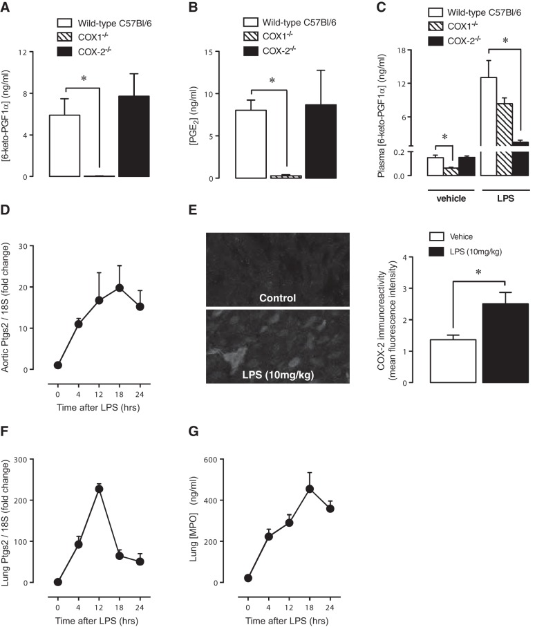
COX-2 expression activity in the vasculature and the effect of LPS. In untreated mice, Ca2+ ionophore-stimulated prostacyclin release (measured as 6-keto-PGF1α) by aortic segments (A) and PGE2 formation in lung homogenates (B) was not altered by COX-2 deletion, but abolished by COX-1 deletion. Accordingly, basal plasma levels of 6-keto-PGF1α were reduced by COX-1 but not COX-2 deletion (C). Administration of LPS (10 mg/kg) increased plasma prostacyclin levels (C), and this effect was COX-2 dependent. LPS also produced a time-dependent increased in aortic COX-2 (Ptgs2) gene expression (D), and this was detectable at 4 h as an increased COX-2-like immunoreactivity (E). LPS also produced a time-dependent increased in lung COX-2 gene expression (F) and neutrophil infiltration into the lung, measured as increased myeloperoxidase levels (G). Data are the means ± se for tissue from n = 4–8 mice aged 10–12 wk. Data were analyzed using unpaired t test (E) or 1-way ANOVA followed by Bonferroni's multiple comparison test (A–C). *P < 0.05.

