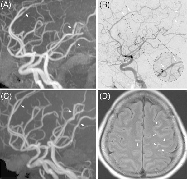
Figure 3.

Imaging findings of reversible cerebral vasoconstriction syndrome. Multifocal vasoconstriction demonstrated by magnetic resonance angiography (MRA) (A) and catheter angiography (B), involving the anterior, middle, and posterior cerebral arteries. Follow-up MRA (C) revealed significant interval resolution of the previous lesions. Axial fluid attenuated inversion recovery (FLAIR) imaging revealed linear hyperintensity lesions in the sulci of the bilateral frontal lobes (D).
