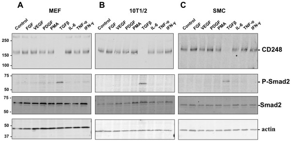
Figure 1.

Expression of CD248 by mesenchymal cells in response to cytokines and growth factors. Murine embryonic fibroblasts (MEF) (A), 10 T1/2 cells (B) and murine aortic smooth muscle cells (SMC) (C) were incubated for 48 hrs with FGF (10 ng/ml), VEGF (20 ng/ml), PDGF (20 ng/ml), PMA (60 ng/ml), TGFβ (3 ng/ml), IL-6 (10 ng/ml), TNF-α (10 ng/ml), or IFN-γ (10 ng/ml). Cells were lysed and separated by SDS-PAGE under non-reducing conditions for Western immunoblotting to detect CD248 and phosphorylated Smad2. Equal loading was confirmed with actin control. Only TGFβ suppressed expression of CD248, while inducing phosphorylation of Smad2. Results are representative of 3 independent experiments. Molecular weight markers in kDa are shown on the left.
