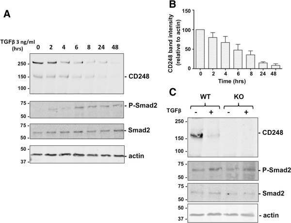
Figure 3.
Temporal response of CD248 to TGFβ. (A) MEF were incubated for 0-48 hrs with TGFβ 3 ng/ml. Expression of CD248 and phosphorylation of Smad2, were detected by Western blot. (B) CD248 expression relative to actin expression was quantified by densitometry (n = 3 experiments) and results were normalized to the no-treatment condition. CD248 expression decreases as Smad2 is phosphorylated. (C) CD248WT/WT (WT) or CD248KO/KO (KO) MEF were exposed to TGFβ (0 or 3 ng/ml) for 48 hrs and lysates were Western blotted. Representative blots from 3 experiments are shown. Smad2 and ERK1/2 are phosphorylated in response to TGFβ even in cells that lack CD248.

