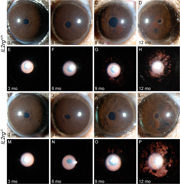
Figure 2.
Reduced NK cell number has no influence on Tyrp1bGpnmbR150Xmediated iris disease. Despite some variability from eye to eye, the range and prevalence of phenotypes at each age was indistinguishable between genotypes. The first row for the indicated genotype shows broad beam illumination to assess the dispersed pigment phenotype and the iris stromal morphology. The second row shows transillumination patterns assaying for the degree of iris depigmentation. The onset and progression of iris disease in B6.D2-GpnmbR150XTyrp1bIl2rg-/- (I to P) is similar to that in B6.D2-GpnmbR150XTyrp1bIl2rg+/+ mice (A-H). Eyes of young B6.D2-GpnmbR150XTyrp1bIl2rg-/- mice exhibit healthy irides (I,M). At 6 mo, mutant mice exhibit characteristic swelling of the peripupillary region (J). At 9 mo the iris becomes atrophic, a transillumination defect is visible, and prominent dispersed pigment is visualized (K,O). At 12 mo, the degree of iris atrophy is prominent as visualized by presence of distinct iris holes and severe transillumination and pigment dispersion (L,P).

