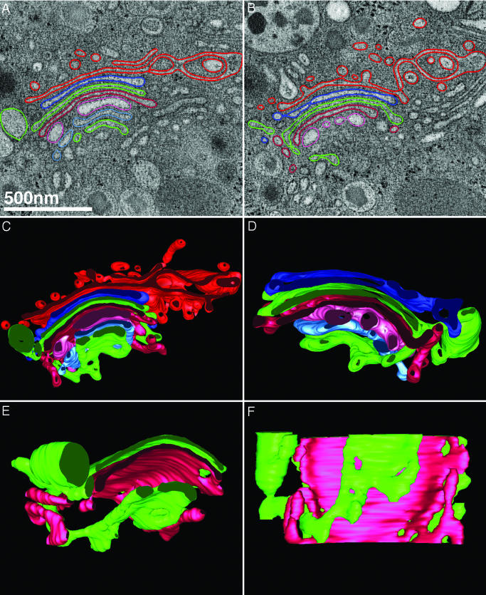
Fig. 3.
Cisternal bypass at the stack periphery where the Golgi ribbon is unbranched. An example of the third type of connection between cisternae at different levels in the Golgi of another islet beta cell following glucose-stimulation (60 ± 15 min) is shown. Neither of the two tomographic slices shown in A and B are able to convincingly demonstrate that the cisternae (green) located at the cis- and trans-sides of the stack (lower and upper faces of the stack, respectively) are directly connected to one another at the periphery of the ribbon. However, when contoured in the z axis and rendered in 3D, the tubular membrane lumen connecting each of the flattened, saccular cisternae can be followed in the context of all cisternae within the stack (C), including the clathrin-coated trans-most cisterna (red) that presents numerous vesicular and tubular profiles. (D) The same stack shown in C but viewed from behind (rotated 180°) and without the trans-most cisterna displayed. In E and F, the connected cisternae (green) are viewed from the front and from below, together with an interceding cisterna (cherry red) for perspective, to allow the reader to visualize the connecting region. In C and D, it should be noted that none of the cisternae in the Golgi stack are simply “folded over”; rather, the connection takes the form of a membrane tubule. Movies 3 and 4 of the tomogram of this region shown in A and B (Movie 3), and of the 3D model data generated by tracing membranes within the reconstructed volume shown in C–F allow the interconnected cisternae to be viewed unambiguously as they are rotated around the x and y axes (Movie 4). Although vesicles are present in the Golgi regions of these cells, we have not shown them for the sake of clarity. However, discrete vesicles associated with the trans-Golgi (red) can be viewed in Movie 4.

