
Figure 1. The Hp UreI urea channel hexamer.
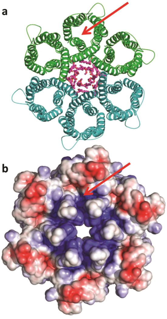
a, Ribbon diagram of hexamer surrounding the lipids of the central bilayer (purple sticks). The C6 hexamer is generated from the three protomers of one asymmetric unit (green) by the crystallographic two-fold axis (second asymmetric unit in teal). b, Electrostatic potential at the periplasmic hexamer surface computed at pH 5.3, the pH at which the crystals were grown (red: -4 kT/e, blue: +4 kT/e). The electrostatic potential was calculated with the program APBSmem22. The green arrow pinpoints the entrance to one of six urea channels.