
Figure 2. Residues lining the channel.
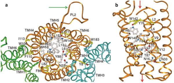
Residues conserved in the AmiS/UreI superfamily (aligned in Supplementary Fig. 11) are clustered in the channel and at the protomer interface. a, View from periplasm showing the conserved residues of protomer A (gold ribbon), and open channel of protomers B & C (green and blue ribbons) when only the protein backbone is shown. For clarity only selected side chains are labeled. Residues conserved in all members of the AmiS/UreI superfamily are white and additional residues conserved in the subset of known urea channels (including HpUreI) are yellow. The arrow points to the proton-sensing periplasmic loop 2 (PL2) of protomer A. b, View parallel to membrane with fully conserved residues (white) clustering in the middle of the channel pore and residues additionally conserved in the urea channels in yellow. The green arrows show the regions of urea entry (top, periplasmic side) and exit (bottom, cytoplasmic side).