
Figure 1.
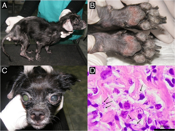
Clinical and histopathological aspects in a 6 year old mixed breed dog with leishmaniasis. A. Muscle waisting of the body muscles, exfoliative dermatitis, difuse alopecia; B. Footpads thickening and scaling, onychogryphosis; C. Nasal depigmentation, blepharitis, anterior uveitis; D. Skin biopsy: few round to oval, 2–3 micron protozoal amastigotes (arrows) located intrahistiocytic or extracellularly. Several small lymphocytes are also present in this section of the superficial dermis (hematoxylin and eosin stain).