Fig. 1.
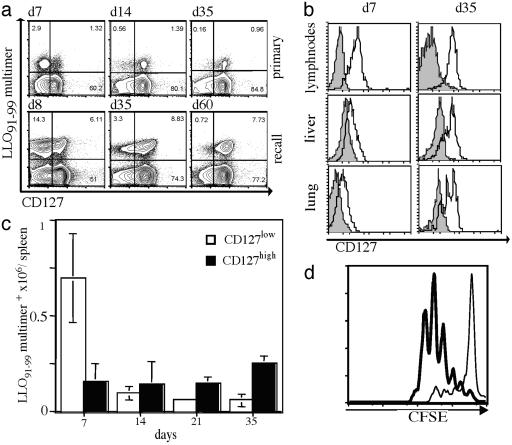
CD127 (IL-7R) surface expression as a marker for memory T cells. (a) A cohort of BALB/c mice was infected with a sublethal dose (≈0.1 × LD50) of WT Lm, followed by secondary infection (≈10 × LD50) 5 wk later. Representative dot plots of splenocytes (gated on live CD8+ T cells) stained with H2-Kd/ LLO91–99 tetramers (y axis) and for CD127 surface expression (x axis) are shown for the indicated time points during primary (Upper) and recall (Lower) responses. (b) Lymphocytes from different organs were isolated during the primary effector (7 days after infection) and memory (35 days after infection) phases. Representative histograms are gated on CD8+ and H2-Kd/ LLO91–99 tetramer-positive cells and show CD127 staining (open) and unstained controls (filled). (c) Kinetics of the absolute numbers (y axis) of CD127 high-(filled) and low (open)-expressing CD8+ and H2-Kd/LLO91–99 tetramer-positive cells; six individual mice per time point. (d) CD127 high- and low-expressing CD8+, H2-Kd/LLO91–99 multimer-positive cells were sorted directly ex vivo 10 days after Listeria infection. Proliferation of carboxy fluorescein succinimidyl ester-labeled cells was analyzed after anti-CD3 stimulation; CD127high (bold line) or CD127low (thin line) cells.

