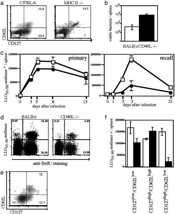
Fig. 4.
Impaired memory responses in MHC II–/– and CD40L–/– mice correlate with early defects in the generation of distinct T cell subsets. (a) WT C57BL/6 (Left) and MHC II-deficient (Right) mice were infected with a sublethal dose of ovalbumin-expressing Lm; dot plots of CD8+, H2-Kb/SIINFEKL-positive T cells stained for CD62L (y axis) and CD127 (x axis) 10 days after primary infection are shown. (b) Numbers of viable bacteria in the spleen 3 days after reinfection (≈10 × LD50; 5 wk after primary infection) with Listeria in CD40L–/– (filled) and WT BALB/c (open); n = 3 per group. (c) Kinetics of LLO91–99-specific T cells determined by H2-Kd/LLO91–99 tetramer staining in CD40L–/– (•) or WT BALB/c mice (□) after primary (0.1 × LD50) and recall infection (2.5 × LD50; 5 wk after primary infection) with Lm; three mice per time point. (d) WT BALB/c mice and CD40L–/– mice received BrdUrd by drinking water during recall infection with Lm (2.5 × LD50; 5 wk after primary infection) during the entire expansion phase (days 1–5); dot plots show BrdUrd incorporation (x axis) in CD8+ T cells stained with H2-Kd/LLO91–99 tetramers (y axis), staining was performed on day 5. (e) Double staining of CD8+, H2-Kd/ LLO91–99 tetramer-positive cells from a CD40L–/– BALB/c mouse for CD62L (y axis) and CD127 (x axis) 10 days after primary infection (WT BALB/c control see Fig. 2c). (f) Absolute numbers of LLO91–99 tetramer-positive T cell subsets in the spleen determined 10 days after primary infection by staining for CD62L and CD127.

