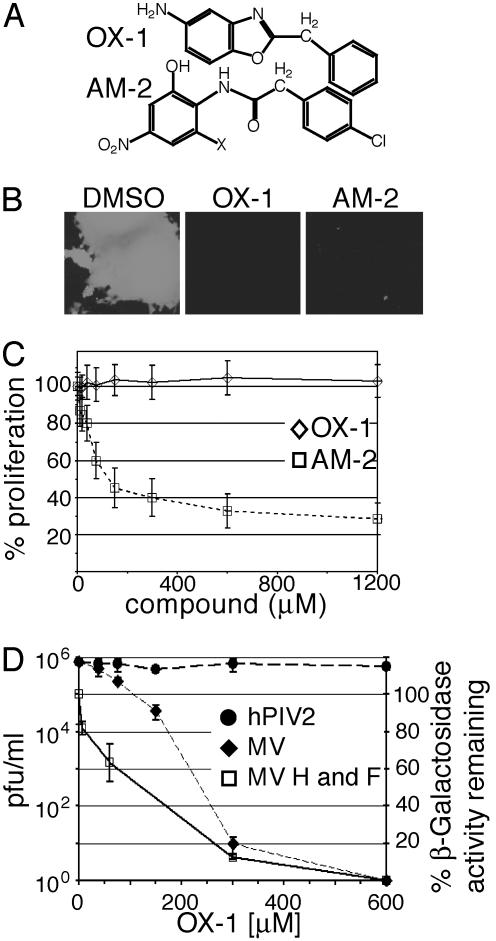
Fig. 1.
Identification of a first-generation lead compound. (A) Structures of compounds OX-1 and AM-2. (B) Inhibitory activity of compounds OX-1 and AM-2. Cells were infected with MV-GFP in the presence of compounds or DMSO and photographed at a magnification of 400×. (C) Proliferation assay of cells incubated in the presence of compound. Values indicate the percentage of signal intensity as compared with cells treated with DMSO. (D) Cells infected with MV-Edm or hPIV2 were incubated in the presence of different OX-1 concentrations as indicated, and virus yields were determined by TCID50 titration (left axis). A β-galactosidase reporter assay was used for quantification of cell fusion mediated by transiently expressed MV glycoproteins in the presence of different OX-1 concentrations; the percentage of β-galactosidase activity as compared with cells treated with DMSO is given (right axis).

