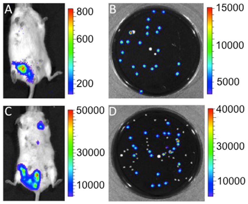
Figure 2. Post-partum L. monocytogenes infection translocates to mammary glands and breast milk.

Mice were inoculated intragastrically with 106 CFU of LM10403S 3 days after parturition. The dams were removed from their pups for two hours to allow for mammary engorgement and injected intraperitoneally with 10 units of oxytocin to stimulate milk letdown to the gland cistern at (A) 72 h and (C) 96 h after infection with L. monocytogenes. Milk was expressed from luminescent glands onto sterile filter paper and placed in BHI media for a 24 h enrichment culture prior to serial dilution in phosphate buffered saline and plating on blood agar. Plates incubated at 37°C for 24 h and were imaged 24 h later. Panel B illustrates post enrichment 10−8 dilution of milk expressed 72 h after infection and Panel D a post enrichment 10−7 dilution of milk expressed 96 h after inoculation. Plates exhibit luminescent L. monocytogenes colonies amongst non-luminescent colonies of bacteria. Pup mortality was not observed in dams infected 72 h post-partum. Results are representative of five experiments with two mice per experiment.
